
PH can be difficult to diagnose in routine medical exams because the most common symptoms of PH — breathlessness, fatigue and dizziness — are associated with many more common conditions such as asthma or chronic obstructive pulmonary disease.
These symptoms include:
- Shortness of breath
- Fatigue
- Fainting or lightheadedness
- Chest pain
- Heart palpitations
- Edema or swelling of the abdomen, legs or feet
Some patients also report cognitive symptoms, such as minor memory issues and “brain fog”. Those issues could be related to problems PH causes in the circulatory system, such as preventing oxygenated blood from freely flowing throughout the body, including to the brain. Symptoms are common across all types of PH.
How PH is diagnosed
When doctors suspect someone has PH, they will:
- Review the patient’s medical and family history
- Perform physical exams
- Perform diagnostic tests
Before conducting any tests, your doctor may perform a physical examination. During the exam, they may look for:
- Visible and/or enlarged veins along the sides of the neck
- Irregular heart sounds
- Swelling in your legs, ankles and abdomen (edema).
After your physical exam, your doctor may then order a series of tests. Diagnostic tests can vary from clinic to clinic. They include testing of pulmonary pressures and function, exercise tolerance tests, blood tests and imaging of the heart.
These tests are categorized as:
PH detection tests
PH is rarely diagnosed through a routine exam and requires specific tests including a right heart catheterization for an accurate diagnosis.
Right heart catheterization
If the results of initial diagnostic tests below point to PH, your doctor will schedule this more involved test. The right heart cath is the only test that directly measures the pressure inside the pulmonary arteries, and it should be done in all patients at least once to confirm a patient’s diagnosis of PH.
Right Heart Cath
Normal pulmonary artery pressure ranges from 8 to 20 mmHg at rest but people with PH will generally have an average resting pulmonary artery pressure above 20 mmHg.
About the RHC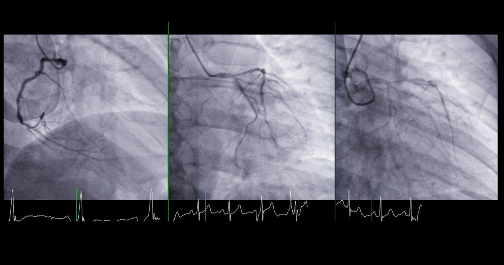
Chest X-rays
A chest X-ray is a non-invasive test that uses radiation to take a picture of the structures inside the chest. Doctors can look at the heart, lungs, blood vessels and bones. When examining patients for PH, doctors will look at the shape of the heart to see if the chambers are larger than expected. They will check whether the pulmonary arteries are more visible than usual. And they will look for signs of lung disease, infection or fluid in the lungs (e.g., pleural effusion).
With chest X-rays, doctors may see evidence of heart disease (Group 2 PH) or lung disease (Group 3 PH).
Electrocardiogram (EKG or ECG)
An electrocardiogram is a simple, painless test that shows the electrical activity of the heart. In most cases, 12 sticky pads are placed on the chest, arms and legs and connected to a machine that prints the electrical activity of the heart.
Doctors use EKGs to check how fast the heart beats, whether the beats are regular, the strength of the electrical pulse in the heart that causes the “beat” and prior heart damage.
Echocardiogram (echo)
An echocardiogram is a painless procedure that takes an ultrasound of the heart.
During an echo, a sticky gel is placed on the chest near the heart, and a technician or doctor will glide a wand connected to a computer over the gel. The wand transmits sound waves to the computer. The computer translates the sound waves into pictures and videos of the heart.
Doctors use the images from the ultrasound to
- Assess the size of the heart
- Determine how well it pumps
- Review how fast the blood moves
- Look at the right side of the heart (the side that pumps blood into the lungs)
Sometimes doctors inject saline or dye into veins during echocardiograms to better see the heart and detect abnormal blood flow. Doctors also use echocardiograms to look for evidence of left heart disease (Group 2 PH), but the tests can’t definitively diagnose PH.
Echocardiograms provide rough estimates of pressure in the pulmonary artery. They estimate only systolic pulmonary artery pressure (when the heart is squeezing), which will be higher than the mean pulmonary artery pressure measured with a right heart cath. The mean pulmonary artery pressure is an average of systolic and diastolic pressures (when the heart relaxes between beats). It will be lower on the right heart cath than an estimated pressure from an echo.
Cardiac magnetic resonance imaging (MRI)
If images from an echocardiogram are unclear, or doctors want more precise measurements of right heart size and function, they might use cardiac MRIs to look at heart function. The procedure provides images of the heart made by a powerful magnetic field and radio waves.
Identifying group, type and severity of PH
Additional tests help identify the type or group of PH as well as a patient’s risk classification. Tests can also help determine optimal treatment options.
Additional TestsRisk assessment
You may hear your care team talk about your risk classification or prognosis. Here’s how that assessment works and what it means.
Risk Assessment in PH